Radiographs
Advanced Dental Imaging For Precise, Detailed Dental Treatments
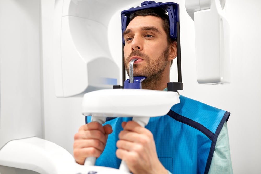
We have an on-site OPG radiograph and a Cone Beam CT scanner to get an exact picture of your teeth and jaws.

Radiographs At Available Dental Care Campbelltown
This radiograph produces images such as:
OPG
OPG radiographs use a panoramic or wide view x-ray of the face, which displays all the teeth of the upper and lower jaw on a single film. It demonstrates the number, position and growth of all the teeth including those that have not yet surfaced or erupted. It is different from the small close up x-rays dentists take of individual teeth.
An OPG may also reveal problems with the jawbone and the joint which connects the jawbone to the head, called the Temporomandibular joint or TMJ. It may be requested for the planning of orthodontic treatment, for assessment of wisdom teeth or for a general overview of the teeth and the bone which supports the teeth.
This is safe and produces minimal radiation

3D and Cone Beam CT scan
A dental cone beam (CB) CT scanner uses x-rays and computer processed x-ray information to produce 3D cross-sectional images of the jaws and teeth.
It is a smaller, faster and safer version of the regular CT scanner.
This will give us detailed information which cannot be obtained from normal x-ray examinations.
Through the use of a cone shaped x-ray beam, the radiation dosage is lower, and the time needed for scanning is reduced.
If you are being considered for dental implants or other special procedures, it enables us at Available Dental Care to assess the exact shape of the bone. These scans can be very helpful in guided implant surgery, high resolution guided endodontics, impacted wisdom surgery and Orthodontics.
BW’s
Bite wing radiographs use x-rays to show details of the upper and lower teeth in one area of the mouth. Each bite wing shows a tooth from its crown (the exposed surface) to the level of the supporting bone.
Bite wing x-rays detect decay between teeth and changes in the thickness of bone caused by gum disease and can also see any wear or breakdown of dental fillings.
Bite wing x-rays can also help determine the proper fit of a crown (a cap that completely encircles a tooth) or other restorations (eg, bridges).

